Obese Face Higher Radiation
Weight★No★More℠ Diet Center
Radiology plays an important role in medical care, but the imaging process becomes more difficult for obese patients. Exams considered simple to perform on average sized patients need to be adapted in order to obtain quality images of obese individuals.
Given the alarming trend of obesity and the long list of associated health risks, it is no surprise that an increasing number of overweight and obese individuals are entering radiology clinics. Most medical imaging equipment, however, is designed with people of an average build in mind and not for overweight and obese patients. As a result, they are exposed to higher levels of radiation during routine X-ray and CT scans.
Back in 2012, researchers at the Rensselaer Polytechnic Institute were the first to calculate exactly how much additional radiation obese patients receive when undergoing a routine CT scan.
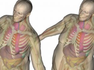
Thick layers of fat (see photo below) make it difficult for the x-ray beam to penetrate the patient. When regular settings on the scanners were set, blurry images were produced.
In order to assure there were enough x-ray photons passing through the body to form a good image, the power (technically referred to as ‘tube potential’) had to be turned up.
The Rensselaer study showed that the internal organs of obese men receive 62% more radiation during a CT scan than those of normal weight men. For obese women, it was an increase of 59%.
The risk associated with a radiation dose from a single CT scan or X-ray is small. However, if you are over-fat, you’re at a greater risk for a long list of medical diagnoses, requiring more imaging . . . and radiation exposure is cumulative over your lifetime.
_______________________________
Additional reading:
Higher Radiation Dose Needed to X-Ray Obese Patients Increases Cancer Risk
Slimcerely yours℠,


